Sleep apnea is a chronic respiratory disorder characterized by repeated pauses in breathing during sleep. Globally, it affects close to one billion individuals, thereby creating a major public health burden1. Although it is often mistaken for harmless snoring, the condition represents a dangerous cycle of suffocation followed by neurological rescue. Each breathing pause lowers blood oxygen levels, which then trigger stress responses throughout the body. Over time, this recurring pattern promotes systemic inflammation, cardiovascular disease, and metabolic dysfunction2.
Modern medicine now frames sleep apnea as a combined failure of airway physics and neural control3. Structural vulnerability allows the airway to collapse, while unstable brain signaling fails to maintain a consistent breathing rhythm. For this reason, researchers increasingly describe the disorder as a systems-level breakdown rather than a single anatomical defect. This integrated view has gradually shifted treatment away from simple airflow support alone4. Instead, therapies increasingly target muscle coordination, neural feedback regulation, and metabolic drivers, and this way long-term outcomes are improving5.
To understand how these diverse mechanisms interact, it is helpful to first examine the primary clinical forms of sleep apnea and the biological differences that distinguish them. These clinical patterns provide the foundation for understanding how airway collapse and neural instability develop during sleep.
Types of sleep apnea and clinical phenotypes
Obstructive and central mechanisms
Sleep apnea appears in several forms, although two dominant mechanisms account for most cases. Understanding these mechanisms helps clinicians identify the biological driver of disease and select appropriate therapies.
Obstructive sleep apnea (OSA) is the most common form and affects nearly 936 million adults aged 30 to 69 worldwide6, 7. It is defined by an apnea–hypopnea index of five or more events per hour. In OSA, the airway collapses physically during sleep, which blocks airflow despite ongoing breathing effort from the chest and diaphragm.
Central sleep apnea (CSA), in contrast, arises from impaired signaling between the brainstem and respiratory muscles8. In CSA, breathing temporarily stops because the brain fails to send appropriate signals to initiate inhalation.
Although these mechanisms differ in origin, the physiological consequences are remarkably similar. Both forms interfere with sleep continuity and cause repeated oxygen drops, which then expose tissues to chronic oxidative stress. Over time, this stress damages blood vessels, disrupts metabolic balance, and increases cardiovascular risk.
Early identification is therefore essential. When clinicians determine the dominant phenotype, they can tailor treatment strategies more precisely, which helps improve both patient adherence and therapeutic outcomes.
Pathophysiology and the physics of negative pressure collapse
Airway mechanics during sleep
To understand why airway obstruction occurs so frequently during sleep, it is necessary to examine the mechanical properties of the upper airway4.
The upper airway functions as a flexible tube that lacks rigid skeletal support. During sleep, muscle tone naturally decreases, which narrows the pharyngeal diameter. As inhalation begins, the chest generates negative pressure to draw air inward. In susceptible individuals, this suction pulls relaxed tissues inward until the airway seals shut. As a result, airflow stops completely even though the body continues attempting to breathe.
This process closely follows the Starling resistor model, which describes collapsible tubes exposed to external pressure9. In this model, airflow becomes limited when external pressure exceeds the internal pressure that keeps the tube open. Once a critical pressure threshold is reached, the airway collapses regardless of how forcefully a person attempts to inhale.
Over time, the brain recognizes increased carbon dioxide concentrations and decreased oxygen levels, and this detection triggers a brief neurological arousal. This response restores muscle tone and reopens the airway. Despite the rapid resumption of breathing, sleep architecture becomes disrupted, resulting in repeated loss of restorative sleep stages.
Physiological consequences of repeated collapse
When airway collapse occurs dozens or even hundreds of times each night, a cascade of physiological responses follows. The sequence below illustrates the typical progression of events during obstructive apnea episodes10.
- Airway narrowing intensifies during deep sleep, which reduces pharyngeal diameter and increases the likelihood of mechanical collapse.
- Negative pressure then exceeds structural support limits, thereby forcing the airway to close abruptly despite continued breathing effort.
- Intermittent hypoxia activates the sympathetic nervous system, and this way the brain rapidly induces a brief awakening that restores breathing.
- Rapid re-oxygenation follows each event, which generates reactive oxygen species that damage vascular endothelium over time.
Because this cycle repeats throughout the night, the cumulative physiological burden becomes substantial.
Central sleep apnea and feedback loop instability
While obstructive apnea results primarily from airway mechanics, CSA reflects a failure of respiratory control.
The brainstem continuously monitors carbon dioxide levels in the blood in order to regulate breathing rhythm. In CSA, this feedback system becomes unstable. Small fluctuations in carbon dioxide trigger exaggerated responses, thereby producing cycles of overbreathing followed by pauses in respiration11.
Engineers describe this instability using the concept of loop gain, which refers to the sensitivity of a feedback system12. When loop gain becomes excessively high, the system overcorrects after small disturbances and begins to oscillate. In biological terms, breathing waxes and wanes instead of remaining steady.
This pattern frequently appears in individuals with heart failure or neurological disease13. Accordingly, CSA illustrates how respiratory instability can emerge from feedback loop physics rather than airway anatomy alone.
The genioglossus muscle and neuromuscular coordination
The tongue as the primary airway stabilizer
Airway stability during sleep does not depend solely on anatomical structure. Neuromuscular coordination also plays a crucial role in maintaining airflow.
The genioglossus muscle, which forms the bulk of the tongue, acts as the primary stabilizer of the upper airway14. During wakefulness, this muscle contracts reflexively to keep the tongue positioned forward. However, muscle activity decreases significantly during sleep, particularly in individuals predisposed to airway obstruction.
As the tongue relaxes, it may fall backward toward the throat, which narrows or blocks the airway. This neuromuscular failure therefore contributes directly to OSA.
Hypoglossal nerve stimulation was developed to address this mechanism15. A small, implanted device delivers timed electrical impulses to the hypoglossal nerve during inspiration. These impulses move the tongue forward, thereby preventing airway collapse.
By restoring coordinated muscle activity, this therapy targets an underlying cause of airway obstruction rather than simply compensating for its consequences.
Clinical presentation and the gender symptom gap
Several physiological and phenomenological patterns help explain how sleep apnea manifests differently across sexes. The following observations summarize key differences identified in clinical studies.
- Early diagnostic frameworks emphasized loud snoring and witnessed apneas, which occur more commonly in men, leading to historical underdiagnosis in women whose symptom patterns often diverge from this classic picture16.
- Male patients often exhibit pronounced daytime sleepiness, which frequently interfere with occupational performance and daily activities.
- Female patients commonly report fragmented sleep, persistent fatigue, mood disturbances, insomnia, or morning headaches17. These symptoms are less obviously linked to a breathing disorder and are often attributed to stress or anxiety, contributing to diagnostic delay. Yet untreated sleep apnea exposes women to the same cardiovascular and metabolic risks observed in men.
- Men tend to accumulate central and neck fat, which increases mechanical pressure on surrounding airway tissues and promotes collapse18.
- Postmenopausal women experience a sharp rise in apnea prevalence, thereby approaching rates observed in men16.
- Women often have shorter apnea events, yet these episodes still produce significant sleep fragmentation and metabolic stress17.
Recognizing these differences is therefore essential for equitable care. Clinicians increasingly evaluate overall symptom burden rather than relying exclusively on “classic” apnea presentations.
Psychological and cognitive consequences of sleep fragmentation
The repeated interruptions that occur during apnea episodes extend beyond breathing disturbances. They also affect brain function significantly.
Each apnea event triggers a brief arousal response that activates the body’s fight-or-flight system19. Thus, patients experience a state of chronic physiological stress throughout the night. Over time, many individuals develop impaired concentration, memory lapses, and slowed reaction times, which collectively appear as persistent brain fog.
Furthermore, deep sleep stages play a critical role in clearing metabolic waste from the brain. When these stages are repeatedly interrupted, neural maintenance processes cannot function efficiently.
Researchers are therefore investigating whether chronic sleep fragmentation contributes to early cognitive decline. In turn, treating airway instability may protect not only sleep quality but also long-term neurological health20.
Metabolic dysfunction and the bidirectional feedback loop
The relationship between apnea and insulin resistance
Sleep apnea is now recognized as a major contributor to metabolic syndrome21. Each apnea episode triggers the release of cortisol and adrenaline, which prompt the liver to release glucose into the bloodstream22. This response provides energy for rapid awakening and emergency breathing restoration.
However, repeated glucose surges gradually reduce insulin sensitivity. As insulin resistance develops, fat storage increases, particularly in visceral tissues and the upper airway.
This anatomical change worsens airway obstruction, thereby creating a self-reinforcing cycle. Weight loss becomes progressively more difficult because hormonal signals begin to favor energy conservation and increased sugar cravings.
Breaking this cycle therefore requires addressing both breathing instability and metabolic regulation simultaneously.
Key metabolic consequences
Several metabolic changes contribute to the long-term health risks associated with untreated sleep apnea.
- Intermittent hypoxia activates sympathetic pathways, thereby elevating blood pressure and heart rate throughout the night10.
- Chronic cortisol exposure promotes visceral fat accumulation, which increases crowding around airway structures23.
- Insulin resistance develops as an adaptive response, yet prolonged resistance eventually contributes to type 2 diabetes24.
- Oxidative stress damages pancreatic tissue, which reduces the body’s capacity to produce insulin efficiently25.
Pharmacological advances in metabolic airway management
Because metabolic dysfunction contributes directly to airway obstruction, pharmacological therapies have recently gained attention as complementary treatments. Recent pharmacological developments have therefore expanded treatment options for obesity-related sleep apnea.
Glucagon-like peptide-1 receptor agonists, including tirzepatide, received regulatory approval in 2024 for obese adults with OSA26. These medications promote substantial weight loss while simultaneously reducing systemic inflammation. As adipose tissue decreases in the tongue and neck, airway diameter increases measurably. Clinical trials have reported reductions in the apnea–hypopnea index of twenty-five to thirty events per hour in some patients.
In certain cases, partial or even complete disease remission has been observed28. This strategy therefore targets the metabolic root of airway collapse, thereby complementing traditional mechanical therapies.
Modern diagnostics and home-based testing
From sleep labs to wearables
Advances in technology have significantly transformed how sleep apnea is diagnosed.
Traditional laboratory sleep studies remain highly accurate, yet they are expensive and often uncomfortable. Modern home sleep testing devices offer a practical alternative28. These compact systems measure oxygen saturation, heart rate, and respiratory effort while patients sleep in their own homes. Because the sleeping environment remains familiar, the recorded data often reflect typical sleep patterns more accurately.
Artificial intelligence (AI) further enhances interpretation by detecting subtle micro-arousals that manual scoring may overlook29. In addition, contactless radar sensors can track chest movement remotely, thereby reducing patient discomfort30.
Emerging diagnostic tools
Several emerging technologies are expanding access to sleep apnea detection and long-term monitoring.
- Wearable rings and adhesive patches measure overnight oxygen levels and heart rate variability, thereby providing early warning signs of sleep-disordered breathing31, 32.
- Contactless radar systems track breathing motion without physical contact, which improves comfort during extended monitoring30.
- AI-based analysis identifies specific collapse patterns, which helps clinicians choose personalized therapies29.
- Home testing platforms significantly reduce diagnostic costs, thereby expanding access for underserved populations28.
- Smartphone-linked monitoring systems provide long-term sleep data, which allows clinicians to track treatment effectiveness over time33.
Advanced therapies and future directions
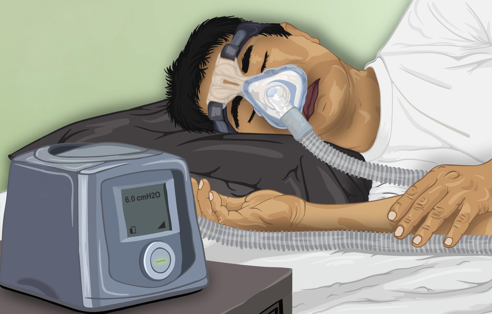
Although continuous positive airway pressure remains highly effective, long-term adherence remains challenging for many patients. As a result, alternative treatments have gained increasing attention.
Oral appliances reposition the jaw forward during sleep, thereby enlarging the airway through mechanical leverage34. In severe cases, maxillomandibular advancement surgery permanently expands airway volume35.
Meanwhile, neuromuscular training approaches are emerging as promising future therapies36. Short daily sessions of targeted electrical stimulation can strengthen airway muscles while patients are awake37. Increased baseline muscle tone reduces nighttime collapse risk, which allows some individuals to maintain stable breathing without masks.
This shift toward physiology-based treatment reflects a broader trend in sleep medicine. As researchers integrate airway mechanics, neural control, and metabolic regulation into a unified framework, clinicians are beginning to match therapies with the specific biological drivers of disease. This approach is making sleep apnea management increasingly precise, personalized, and sustainable.