FAQs
1. Is an ultrasound procedure painful or uncomfortable?
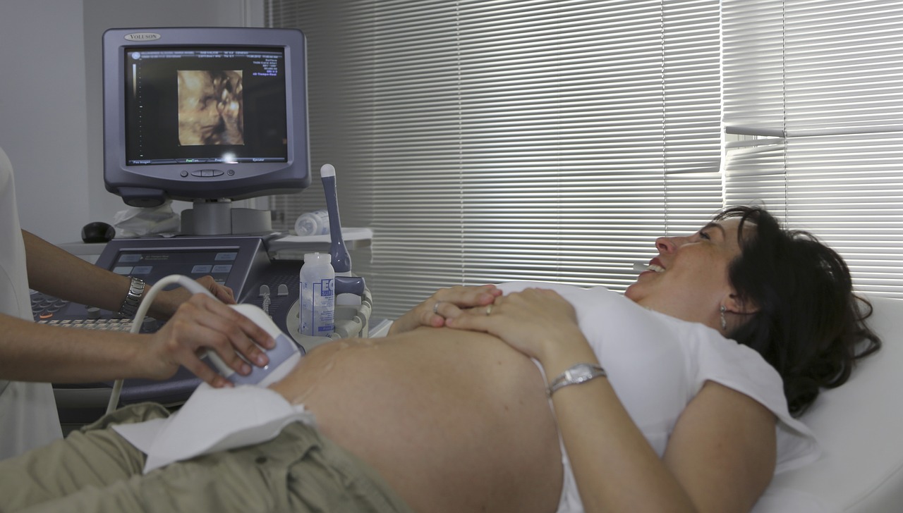
Generally, ultrasound procedures are not painful. You might feel some pressure as the sonographer moves the transducer, especially when imaging deeper organs, but a water-based gel is used to help the transducer glide smoothly.
2. Are there any side effects or risks associated with diagnostic ultrasound?
Diagnostic ultrasound is considered one of the safest medical imaging modalities available. It doesn’t use ionizing radiation, and the sound waves used are at very low power and do not cause any known harmful effects to the body, even for sensitive populations like pregnant women and children. The gel used is hypoallergenic, meaning it is designed to minimize the risk of allergic reaction.
3. What factors can affect the clarity or quality of an ultrasound image?
Several factors can influence the clarity of an ultrasound image, including the presence of excess body fat or significant muscle, and the presence of gas or air, such as in the bowel or lungs. Additionally, the skill and experience of the sonographer play a significant role.
Reference
1. Kasban, H., El-Bendary, M. A. M., & Salama, D. H. (2015). A comparative study of medical imaging techniques. International Journal of Information Science and Intelligent System, 4(2), 37-58.
2. Goldberg, R. L., & Smith, S. W. (2003). Transducers. In Biomedical Imaging, CRC Press. 12-1.
3. Solberg, O. V., Lindseth, F., Torp, H., et al. (2007). Freehand 3D ultrasound reconstruction algorithms—a review. Ultrasound in medicine & biology, 33(7), 991-1009.
4. Douville, N. J., & Bradford, C. R. (2013). Comparison of ultrasound‐guided core biopsy versus fine‐needle aspiration biopsy in the evaluation of salivary gland lesions. Head & neck, 35(11), 1657-1661.
5. Zell, K., Sperl, J. I., Vogel, M. W., et al. (2007). Acoustical properties of selected tissue phantom materials for ultrasound imaging. Physics in Medicine & Biology, 52(20), N475.
6. Liu, H., Wang, J., Zhao, Y., et al. (2021). Doppler ultrasound and contrast-enhanced ultrasound in detection of stent stenosis after iliac vein stenting. BMC Cardiovascular Disorders, 21, 1-7.
7. Drukker, L., Noble, J. A., & Papageorghiou, A. T. (2020). Introduction to artificial intelligence in ultrasound imaging in obstetrics and gynecology. Ultrasound in Obstetrics & Gynecology, 56(4), 498-505.
8. Fadel, B. M., Mohty, D., Kazzi, B. E., et al. (2021). Ultrasound imaging of the abdominal aorta: a comprehensive review. Journal of the American Society of Echocardiography, 34(11), 1119-1136.
9. García-Rivera, E., Cenizo-Revuelta, N., Ibáñez-Maraña, M. A., et al. (2021). Doppler ultrasound as a unique diagnosis test in peripheral arterial disease. Annals of Vascular Surgery, 73, 205-210.
10. Kendall, J. L., Hoffenberg, S. R., & Smith, R. S. (2007). History of emergency and critical care ultrasound: the evolution of a new imaging paradigm. Critical care medicine, 35(5), S126-S130.
11. Maruszczak, K., Kochman, M., Madej, T., et al. (2024). Ultrasound Imaging in Diagnosis and Management of Lower Limb Injuries: A Comprehensive Review. Medical Science Monitor: International Medical Journal of Experimental and Clinical Research, 30, e945413-1.
12. Andersen, C. A., Holden, S., Vela, J., et al. (2019). Point-of-care ultrasound in general practice: a systematic review. The Annals of Family Medicine, 17(1), 61-69.
13. Kwon, S. H., & Gopal, A. S. (2017). 3D and 4D ultrasound: Current progress and future perspectives. Current Cardiovascular Imaging Reports, 10, 1-13.
14. Garra, B. S. (2007). Imaging and estimation of tissue elasticity by ultrasound. Ultrasound quarterly, 23(4), 255-268.
15. Xie, F., Zhang, D., Cheng, L., et al. (2015). Intradermal microbubbles and contrast-enhanced ultrasound (CEUS) is a feasible approach for sentinel lymph node identification in early-stage breast cancer. World journal of surgical oncology, 13, 1-8.
16. Lee, M. W. (2014). Fusion imaging of real-time ultrasonography with CT or MRI for hepatic intervention. Ultrasonography, 33(4), 227.
17. Song, P., Rubin, J. M., & Lowerison, M. R. (2023). Super-resolution ultrasound microvascular imaging: Is it ready for clinical use?. Zeitschrift für Medizinische Physik, 33(3), 309-323.
18. Manzoor, I., Bacha, R., & Gilani, S. A. (2021). Applications of high-intensity focused ultrasound in the treatment of different pathologies. Journal of Diagnostic Medical Sonography, 37(2), 171-178.
19. Burgess, A., & Hynynen, K. (2014). Drug delivery across the blood–brain barrier using focused ultrasound. Expert opinion on drug delivery, 11(5), 711-721.
20. Xu, Z., Hall, T. L., Vlaisavljevich, E., et al. (2021). Histotripsy: the first noninvasive, non-ionizing, non-thermal ablation technique based on ultrasound. International Journal of Hyperthermia, 38(1), 561-575.
21. Kubanek, J. (2018). Neuromodulation with transcranial focused ultrasound. Neurosurgical focus, 44(2), E14.
22. Ottakath, N., Al-Maadeed, S., Bouridane, A., Chowdhury, M. E., & Sadasivuni, K. K. (2024, March). Wearable Ultrasound devices for continuous health monitoring: Current and future Prospects. In 2024 IEEE 8th Energy Conference (ENERGYCON) (pp. 1-6). IEEE.
23. Wells, P. N. (1999). Ultrasonic imaging of the human body. Reports on progress in physics, 62(5), 671.
24. Quien, M. M., & Saric, M. (2018). Ultrasound imaging artifacts: How to recognize them and how to avoid them. Echocardiography, 35(9), 1388-1401.
25. Pedersen, P. C., Dickson, B. W., & Chakareski, J. (2009, October). Telemedicine applications of mobile ultrasound. In 2009 IEEE International Workshop on Multimedia Signal Processing (pp. 1-6). IEEE.