FAQs
1. How is Mass spectrometry imaging (MSI) used in pharmaceutical development to track drug distribution?
MSI allows researchers to label-free map the precise spatial distribution and concentration of an administered drug and its various metabolites directly within a tissue section. This is vital for pharmacokinetics, as it reveals how a compound penetrates tumors, crosses the blood-brain barrier, or accumulates in organs.
2. What is the fundamental difference in the information provided by FTIR and Raman spectroscopy in biological samples?
FTIR measures light absorption caused by changes in the molecule’s dipole moment (e.g., polar bonds like C=O, N-H). Raman measures inelastic scattering based on changes in molecular polarizability (e.g., non-polar C-C, C=C bonds). This makes Raman highly advantageous for aqueous samples as it is minimally sensitive to water, whereas water strongly absorbs in FTIR.
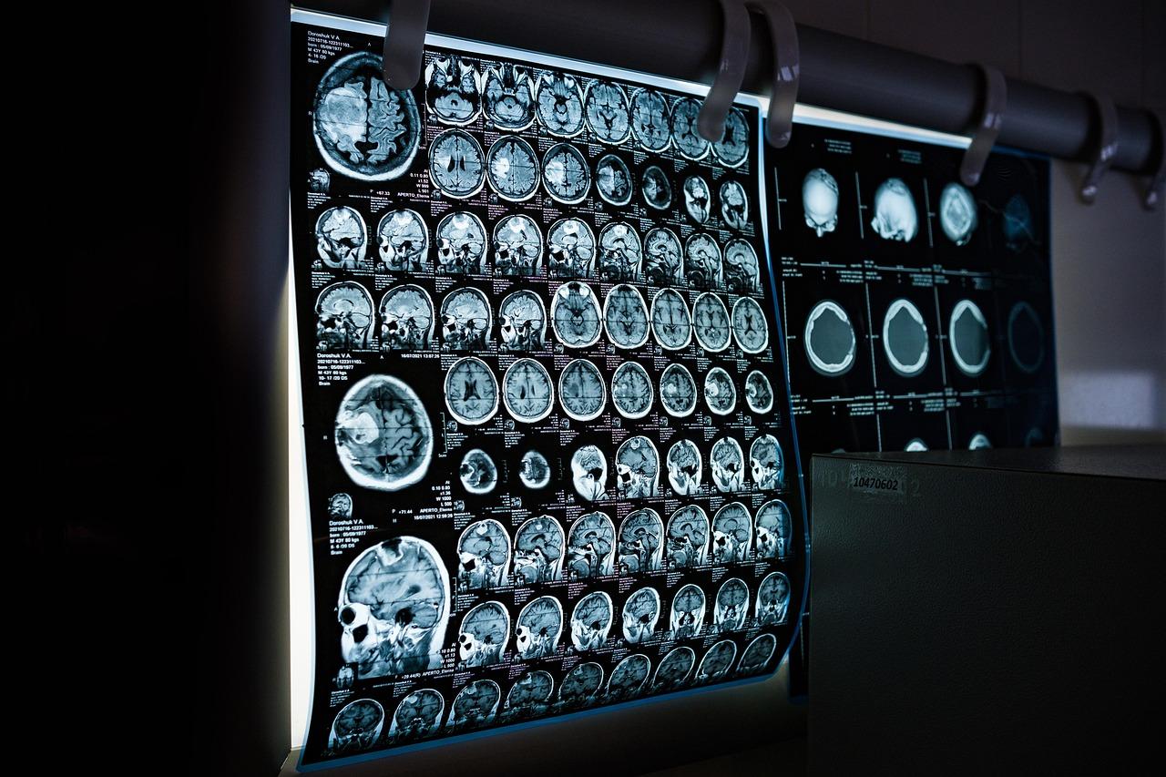
3. How does Functional MRI (fMRI) use the BOLD contrast to map brain activity in cognitive neuroscience?
fMRI maps brain activity by detecting changes in blood oxygenation, known as the blood-oxygenation-level dependent (BOLD) contrast. When a brain region becomes active, blood flow to that area increases, delivering more oxygenated blood than the local neurons consume. Oxygenated and deoxygenated hemoglobin have different magnetic properties, which the MRI scanner detects to indirectly map the areas of increased neural activity.
Reference
1. Zahra, A., Qureshi, R., Sajjad, M., et al. (2024). Current advances in imaging spectroscopy and its state-of-the-art applications. Expert Systems with Applications, 238, 122172.
2. Ferré, G., & Eddy, M. T. (2020). Structural biology of human GPCR drugs and endogenous ligands-insights from NMR spectroscopy. Methods, 180, 79-88.
3. Pence, I., & Mahadevan-Jansen, A. (2016). Clinical instrumentation and applications of Raman spectroscopy. Chemical Society Reviews, 45(7), 1958-1979.
4. Banwell, C. N., & McCash, E. M. (1994). Fundamentals of molecular spectroscopy. Fourth Edition. McGraw-Hill.
5. Boustany, N. N., Boppart, S. A., & Backman, V. (2010). Microscopic imaging and spectroscopy with scattered light. Annual review of biomedical engineering, 12(1), 285-314.
6. Ameh, E. S. (2019). A review of basic crystallography and x-ray diffraction applications. The international journal of advanced manufacturing technology, 105(7), 3289-3302.
7. Tiernan, H., Byrne, B., & Kazarian, S. G. (2020). ATR-FTIR spectroscopy and spectroscopic imaging for the analysis of biopharmaceuticals. Spectrochimica Acta Part A: Molecular and Biomolecular Spectroscopy, 241, 118636.
8. Krishna, R., & Colak, I. (2023). Advances in biomedical applications of Raman microscopy and data processing: a mini review. Analytical Letters, 56(4), 576-617.
9. Van Velzen, M. J. M., Derks, S., Van Grieken, N. C. T., et al. (2020). MSI as a predictive factor for treatment outcome of gastroesophageal adenocarcinoma. Cancer treatment reviews, 86, 102024.
10. Dimakopoulou-Papazoglou, D., Ploskas, N., Serrano, S., et al. (2023). Application of UV–Vis spectroscopy for the detection of adulteration in Mediterranean honeys. European Food Research and Technology, 249(12), 3043-3053.
11. Dowling, R. C., Carroll, G. T., Kirschman, D. L., et al. (2023). Circular dichroism and UV–Vis detection of UV‐induced damage to nucleic acids. Chirality, 35(12), 973-982.
12. Adegoke, J. A., De Paoli, A., Afara, I. O., et al. (2021). Ultraviolet/visible and near-infrared dual spectroscopic method for detection and quantification of low-level malaria parasitemia in whole blood. Analytical Chemistry, 93(39), 13302-13310.
13. Harroun, S. G., & Vallée‐Bélisle, A. (2023). Methods to Characterise Enzyme Kinetics with Biological and Medicinal Substrates: The Case of Alkaline Phosphatase. Chemistry‐Methods, 3(8), e202200067.
14. Drieschner, T., Ostertag, E., Boldrini, B., et al. (2020). Direct optical detection of cell density and viability of mammalian cells by means of UV/VIS spectroscopy. Analytical and Bioanalytical Chemistry, 412(14), 3359-3371.
15. Francis, A. T., Shears, M. J., Murphy, S. C., et al. (2020). Direct quantification of single red blood cell hemoglobin concentration with multiphoton microscopy. Analytical Chemistry, 92(18), 12235-12241.
16. Chen, H., Liu, L., Qian, K., et al. (2022). Bioinspired large Stokes shift small molecular dyes for biomedical fluorescence imaging. Science Advances, 8(31), eabo3289.
17. Elliott, A. D. (2020). Confocal microscopy: principles and modern practices. Current protocols in cytometry, 92(1), e68.
18. McAleer, S., Fast, A., Xue, Y., et al. (2021). Deep Learning–Assisted Multiphoton Microscopy to Reduce Light Exposure and Expedite Imaging in Tissues With High and Low Light Sensitivity. Translational Vision Science & Technology, 10(12), 30.
19. Codron, P., Letournel, F., Marty, S., et al. (2021). STochastic Optical Reconstruction Microscopy (STORM) reveals the nanoscale organization of pathological aggregates in human brain. Neuropathology and applied neurobiology, 47(1), 127-142.
20. Xie, L., Dong, P., Chen, X., et al. (2020). 3D ATAC-PALM: super-resolution imaging of the accessible genome. Nature methods, 17(4), 430-436.
21. Calovi, S., Soria, F. N., & Tønnesen, J. (2021). Super-resolution STED microscopy in live brain tissue. Neurobiology of Disease, 156, 105420.
22. Zou, F., & Bai, L. (2019). Using time-lapse fluorescence microscopy to study gene regulation. Methods, 159, 138-145.
23. Sekar, R. B., & Periasamy, A. (2003). Fluorescence resonance energy transfer (FRET) microscopy imaging of live cell protein localizations. The Journal of cell biology, 160(5), 629.
24. Peled‐Zehavi, H., & Gal, A. (2021). Exploring intracellular ion pools in coccolithophores using live‐cell imaging. Advanced biology, 5(6), 2000296.
25. Ozaki, Y. (2021). Infrared spectroscopy—Mid-infrared, near-infrared, and far-infrared/terahertz spectroscopy. Analytical Sciences, 37(9), 1193-1212.
26. Levin, I. W., & Bhargava, R. (2005). Fourier transform infrared vibrational spectroscopic imaging: integrating microscopy and molecular recognition. Annu. Rev. Phys. Chem., 56(1), 429-474.
27. Lewis, P. D., Lewis, K. E., Ghosal, R., et al. (2010). Evaluation of FTIR spectroscopy as a diagnostic tool for lung cancer using sputum. BMC cancer, 10(1), 640.
28. Krishna, R., & Colak, I. (2023). Advances in biomedical applications of Raman microscopy and data processing: a mini review. Analytical Letters, 56(4), 576-617.
29. Antonio, K. A., & Schultz, Z. D. (2014). Advances in biomedical Raman microscopy. Analytical chemistry, 86(1), 30-46.
30. Aaboubout, Y., Soares, M. R. N., Schut, T. C. B., et al. (2023). Intraoperative assessment of resection margins by Raman spectroscopy to guide oral cancer surgery. Analyst, 148(17), 4116-4126.
31. Ando, M., Sugiyama, K., Kubo, K., et al. (2023). Single-Cell Level Raman Molecular Profiling Reveals the Classification of Growth Phases of Chaetoceros tenuissimus. The Journal of Physical Chemistry B, 127(22), 5027-5033.
32. LaLone, V., Smith, D., Diaz-Espinosa, J., et al. (2023). Quantitative Raman Chemical Imaging of Intracellular Drug-Membrane Aggregates and Small Molecule Drug Precipitates In Cytoplasmic Organelles. Advanced drug delivery reviews, 202, 115107.
33. Schlücker, S. (2014). Surface‐enhanced Raman spectroscopy: concepts and chemical applications. Angewandte Chemie International Edition, 53(19), 4756-4795.
34. Marion, D. (2013). An introduction to biological NMR spectroscopy. Molecular & Cellular Proteomics, 12(11), 3006-3025.
35. Logothetis, N. K. (2008). What we can do and what we cannot do with fMRI. Nature, 453(7197), 869-878.
36. Ranzenberger, L. R., Das, J. M., & Snyder, T. (2023). Diffusion tensor imaging. In StatPearls [Internet]. StatPearls Publishing.
37. Tognarelli, J. M., Dawood, M., Shariff, M. I., et al. (2015). Magnetic resonance spectroscopy: principles and techniques: lessons for clinicians. Journal of clinical and experimental hepatology, 5(4), 320-328.
38. Gross, J. H. (2006). Mass spectrometry: a textbook. Springer Science & Business Media.
39. Aichler, M., & Walch, A. (2015). MALDI Imaging mass spectrometry: current frontiers and perspectives in pathology research and practice. Laboratory investigation, 95(4), 422-431.
40. Garza, K. Y., Feider, C. L., Klein, D. R., et al. (2018). Desorption electrospray ionization mass spectrometry imaging of proteins directly from biological tissue sections. Analytical chemistry, 90(13), 7785-7789.
41. Spruill, M. L., Maletic-Savatic, M., Martin, H., et al. (2022). Spatial analysis of drug absorption, distribution, metabolism, and toxicology using mass spectrometry imaging. Biochemical pharmacology, 201, 115080.
42. Duncan, K. D., Pětrošová, H., Lum, J. J., et al. (2024). Mass spectrometry imaging methods for visualizing tumor heterogeneity. Current opinion in biotechnology, 86, 103068.
43. Scott, A. J., Jones, J. W., Orschell, C. M., et al. (2014). Mass Spectrometry Imaging Enriches Biomarker Discovery Approaches with Candidate Mapping. Health physics, 106(1), 120.
44. Bunaciu, A. A., UdriŞTioiu, E. G., & Aboul-Enein, H. Y. (2015). X-ray diffraction: instrumentation and applications. Critical reviews in analytical chemistry, 45(4), 289-299.
45. Yasaka, K., Akai, H., Kunimatsu, A., et al. (2020). Prediction of bone mineral density from computed tomography: application of deep learning with a convolutional neural network. European radiology, 30(6), 3549-3557.
46. Duncan, K. E., Czymmek, K. J., Jiang, N., et al. (2021). X-ray microscopy enables multiscale high-resolution 3D imaging of plant cells, tissues, and organs. Plant Physiology, 188(2), 831.
47. Triche, B. L., Nelson Jr, J. T., McGill, N. S., et al. (2019). Recognizing and minimizing artifacts at CT, MRI, US, and molecular imaging. RadioGraphics, 39(4), 1017-1018.
48. Zhang, H., Zhang, J., Yuan, C., et al. (2024). Recent advances in mass spectrometry imaging combined with artificial intelligence for spatially clarifying molecular profiles: Toward biomedical applications. TrAC Trends in Analytical Chemistry, 178, 117834.
49. Saba, L., & d’Aloja, E. (2025). Predictive techniques in medical imaging: opportunities, limitations, and ethical-economic challenges. npj Digital Medicine, 8(1), 392.
50. Smith, R., Wright, K. L., & Ashton, L. (2016). Raman spectroscopy: an evolving technique for live cell studies. Analyst, 141(12), 3590-3600.
51. Jung, J. H., Choi, Y., & Im, K. C. (2016). PET/MRI: technical challenges and recent advances. Nuclear medicine and molecular imaging, 50(1), 3-12.
52. Jeong, S., Kim, Y. I., Kang, H., et al. (2015). Fluorescence-Raman dual modal endoscopic system for multiplexed molecular diagnostics. Scientific reports, 5(1), 9455.
53. Liu, Y., Chen, S., Xiong, X., et al. (2025). Artificial intelligence guided Raman spectroscopy in biomedicine: Applications and prospects. Journal of Pharmaceutical Analysis, 15(11), 101271.
54. Frija, G., Salama, D. H., Kawooya, M. G., et al. (2023). A paradigm shift in point-of-care imaging in low-income and middle-income countries. EClinicalMedicine, 62.
55. Kang, D., Li, R., Cao, S., et al. (2021). Nonlinear optical microscopies: Physical principle and applications. Applied Spectroscopy Reviews, 56(1), 52-66.
56. Kelkar, S. S., & Reineke, T. M. (2011). Theranostics: combining imaging and therapy. Bioconjugate chemistry, 22(10), 1879-1903.
57. Ghasemi, M., Nabipour, I., Omrani, A., et al. (2016). Precision medicine and molecular imaging: new targeted approaches toward cancer therapeutic and diagnosis. American journal of nuclear medicine and molecular imaging, 6(6), 310.