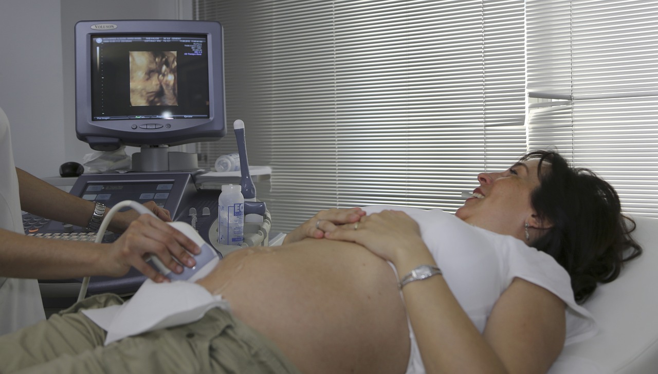
In the rapidly evolving era of modern medicine, diagnostic tools allow healthcare professionals to focus on ailments, continuous health assessment, and the delivery of effective treatments. This includes simple instruments as well as sophisticated machines, which are necessary tools for accurate and timely medical intervention. Ultrasound imaging is one of these diagnostic tools that has played a key role in medical diagnosis, particularly in the fields of obstetrics, cardiology, and abdominal imaging.
It bears several fundamental advantages over other diagnostic techniques, accounting for its widespread use1. Firstly, it uses high-frequency sound waves, in contrast to harmful X-ray radiation, for diagnosis, making it safer for sensitive populations like pregnant women or children. Secondly, it provides real-time images of organs, tissues, and blood vessels, offering information regarding organ movements and blood flow, and is ideal for biopsies and regional anesthesia. Thirdly, it is a cost-effective and portable option, in contrast to expensive diagnostic tools such as MRI/CT, especially in resource-limited healthcare settings, e.g., smaller clinics, remote areas, emergency rooms, or even in ambulances or at patient bedsides.
Established principles of ultrasound imaging
Ultrasound imaging uses high-frequency (2-20 MHz) sound waves to produce images of internal structures by reflection (echo) of these waves from body tissues. The key components of the ultrasound system include a transducer, which transmits the sound waves and captures the returning echoes, and a computer that processes the data to form an image.
Transducer
These are piezoelectric crystals that vibrate upon passage of electric current through them, generating sound waves2. When these sound waves hit tissues or a boundary between different tissues—such as the transition from muscle to bone or fluid—they reflect the waves to the transducer. The time delay between the incident wave and the return of the echoes, and their intensities, are used to calculate the depth and composition of the structure being examined. The returned echoes are converted into electrical impulses by the transducer crystals and are further processed to form the ultrasound image presented on the screen.
Different types of transducers are designed for specific applications:
Linear array transducers
They comprise a straight-line alignment of piezoelectric materials to produce a trapezoidal or rectangular-shaped image. They usually operate at high frequencies ranging from 5-15 MHz or even up to 35 MHz (ref). These transducers are suitable for obtaining high-resolution images for superficial structures such as blood vessels, musculoskeletal structures, breasts, thyroid, etc.
Curvilinear (convex) array transducers
They comprise curved transducers arranged in an arc, producing a wide, fan-shaped image of the objects scanned. They usually operate at low frequencies ranging from 2-6 MHz or even up to 8 MHz (ref). These transducers are suitable for obtaining high-resolution images for deeper structures such as the abdomen, pelvis, and for general abdominal and obstetric examinations, etc.
Phased array transducers
They electronically steer the ultrasound beam by varying the timing of pulses from individual elements, resulting in a triangular-shaped image. They use lower frequencies ranging from 1 MHz to 5 MHz, and sometimes up to 8 MHz. They are ideal for imaging organs that are difficult to access, such as the heart, and for transcranial Doppler (TCD) studies of the brain. They are also used for some abdominal and obstetric applications where a wider field of view from a small acoustic window is needed.
Propagation of sound waves and echoes
The propagation of sound waves depends on the medium they pass through and also on their acoustic impedances that affect how much sound is reflected (echoes) to the transducer. The reflected echoes from different density samples are proportional to the difference in impedances. If the difference in density is increased, the proportion of echo is increased, and the proportion of transmitted sound is proportionately decreased. Echoes are not produced if there is no difference within a tissue or between tissues (appearing dark on the ultrasound image). Homogeneous fluids like blood, bile, urine, contents of simple cysts, ascites, and pleural effusion are seen as echo-free structures. In contrast, denser tissues, like bone or calcifications, reflect more sound and appear bright on the scan.
Image Processing
The computer processes the reflected echoes to create a two-dimensional grayscale image of the internal structures. In modern ultrasound machines, sophisticated software algorithms can generate three-dimensional reconstructions, allowing for more detailed assessments3.
Core diagnostic capabilities of ultrasound imaging
Real-time imaging
Ultrasound imaging provides a dynamic, real-time view of what’s happening inside the body. Examples include systolic and diastolic movements of the heart for diagnosing various cardiac conditions, quantification of blood flow within vessels for detecting blockages, fetal movements, intestinal contractions, and needle guidance during procedures like biopsies or fluid aspirations4.
Anatomical visualization
The difference in the acoustic properties of different types of tissues helps in the anatomical visualization of solid organs like the liver, kidneys, spleen, pancreas, thyroid, and reproductive organs for their size, shape, texture, or abnormal growth of masses, musculoskeletal components, and glands such as the thyroid, salivary glands, and lymph nodes5.
Doppler ultrasound
Doppler ultrasound is an application of ultrasound imaging that provides critical information about moving substances. The phenomenon of a change in the frequency of a wave (sound or light) as received by an observer moving relative to the wave’s source is termed the Doppler effect.
In Doppler ultrasound, the transducer is the source that emits ultrasound that travels through our body and hits moving objects, such as red blood cells. As the blood cells move, they reflect the sound waves to the transducer. If the blood cells are moving towards the transducer, the frequency of the reflected sound waves (echoes) will be higher than the emitted frequency and vice versa. The detector detects this change in frequency (the “Doppler shift”) that is directly proportional to the velocity of the moving blood cells.
Healthcare professionals can decipher critical information from Doppler ultrasound. Examples include assessing blood flow direction, velocity, and character, such as identifying stenosis (narrowing of a blood vessel), thrombus (blood clot), or changes in blood perfusion in tumors6.
Broad clinical applications of ultrasound imaging
Ultrasound is not a niche technology but rather a fundamental diagnostic imaging modality integrated into numerous medical fields due to its versatility, safety (non-ionizing radiation), and real-time capabilities.
Obstetrics and gynecology
Ultrasound is crucial throughout pregnancy for critical monitoring of fetal growth, development, well-being, and detection of congenital anomalies, including uterine fibroids and ovarian cysts7.
Cardiology
This includes assessment of ejection fraction, detection of stenosis, and pericardial effusion6.
Abdominal imaging
Ultrasound is widely used to visualize and assess organs within the abdominal cavity8: for detecting lesions (cysts, tumors); assessing for fatty liver, cirrhosis, or inflammation; identifying gallstones, inflammation (cholecystitis), or polyps; and detecting kidney stones, hydronephrosis (swelling due to urine backup), cysts, or tumors.
Vascular imaging
This includes assessing atherosclerosis for stroke occurrence, diagnosing Deep Vein Thrombosis, and evaluating peripheral artery disease (PAD) by assessing blood flow and blockages in the arteries of the limbs9.
Emergency medicine and critical care
This includes a rapid ultrasound scan performed in trauma patients to quickly identify free fluid (likely blood) indicating internal bleeding that requires immediate intervention10. This involves real-time guidance of needle placement and monitoring for procedures such as central line insertion (placing catheters into large veins); paracentesis (removing fluid from the abdomen); thoracentesis (removing fluid from around the lungs); nerve blockages; and drainage of abscesses or fluid collections.
Musculoskeletal
This involves real-time imaging of tendon or ligament injuries, muscle injuries, and joint effusions11.
The emerging frontier in ultrasound imaging
With the amalgamation of artificial intelligence and modern technological advancements, ultrasound imaging expands its diagnostic scope.
Technological advancements
- The most visible innovation is the remarkable reduction in device size, exemplified by handheld devices connected to smartphones/tablets, and the emergence of point-of-care ultrasound (POCUS)12. This has led to the democratization of imaging access, improved bedside diagnostics, expanded imaging access to remote clinics, and improved physical examinations at primary care settings.
- Technological advancements involve better image resolution with higher-frequency transducers that provide wider bandwidths and advanced array designs. This includes sophisticated algorithms for noise reduction, artifact suppression, and better image clarity. For finer anatomical views, 3D and 4D ultrasound are being used nowadays that offer comprehensive anatomical insights and real-time three-dimensional visualization of dynamic processes13.
Advanced diagnostic techniques
- Elastography: This involves non-invasive measurements of tissue stiffness (elasticity) for diagnosing diseases such as cancer or fibrosis, and for characterizing lesions in the breast or prostate14. The principle is based on tissue deformation under pressure or propagation of shear waves. The stiffer a tissue is, the more it indicates pathology.
- Contrast-enhanced ultrasound (CEUS): This works on the principle of intravenous injection of microscopic gas bubbles (microbubbles)15. These microbubbles enhance vascular signals and provide detailed perfusion information, allowing for more precise detection and characterization of tumors, detailed assessment of myocardial perfusion, and blood flow studies in complex lesions.
- Fusion imaging: This technique involves merging real-time ultrasound images with previously acquired CT/MRI scans, allowing clinicians to combine ultrasound feedback with the comprehensive anatomical context of other modalities16. This leads to highly accurate guidance for biopsies and interventional procedures and is particularly useful in organs with poor ultrasound visibility.
- Super-resolution ultrasound: This is an emerging technique for imaging microvascularization at a resolution beyond conventional limits17.
Therapeutic ultrasound
- High-intensity focused ultrasound (HIFU): This non-invasive method involves focusing high-energy sound waves at a precise focal point to generate localized heat (thermal ablation) and destroy tumors. It is also useful for uterine fibroids, prostate cancer, and even essential tremors (a non-invasive brain therapy)18.
- Focused ultrasound and targeted drug delivery: This technique involves using ultrasound waves to enhance drug delivery at target sites (sometimes crossing the blood-brain barrier) by transiently permeabilizing cell membranes or blood vessels19.
- Histotripsy: An exciting non-thermal approach, histotripsy uses very short, high-energy ultrasound pulses to mechanically liquefy and destroy tissue (e.g., tumors) through cavitation, offering a novel alternative to heat-based ablation20.
- Neuromodulation: Emerging research investigates the use of ultrasound to stimulate or inhibit neural activity in the brain, with studies exploring its role in neurodegeneration21.
Integration with artificial intelligence and machine learning
Integration of artificial intelligence (AI) and machine learning in ultrasound imaging will surely enhance its capabilities and accessibility boundlessly. AI algorithms can improve noise reduction, artifact removal, and overall image quality. AI can assist clinicians in detecting abnormalities and improving diagnostic accuracy by automating complex measurements (e.g., cardiac ejection fraction, tumor volume, fetal biometry)7. AI can perform workflow optimization by automating repetitive tasks, thus improving efficiency. It can also provide feedback on probe positioning and image optimization for novice users, thereby offering real-time guidelines during scans.
Novel applications and research directions
The quest for new domains in ultrasound imaging has led to many pioneering applications such as wearable ultrasound devices for non-invasive continuous monitoring of organ, blood, or other physiological parameters22. Additionally, a few preliminary works also hint at the therapeutic potential for modulating brain activity non-invasively.
Challenges and the future outlook
Despite being widely used for its remarkable advancements, ultrasound imaging still faces certain challenges. One of the most significant limitations is its operator-dependency, requiring skill and experience from the sonographer. Also, attenuation of sound waves by bone and gas creates “blind spots,” restricting its utility in certain specific areas of the body23. Additionally, complications in image interpretation can also occur from artifacts originating during signal reception24. However, the future of ultrasound is continuously developing. The integration of AI will not only reduce operator dependence but also standardize image acquisition and help in interpretation. Additionally, innovations in transducer technology, along with the development of novel contrast agents, smart probes, and integrated sensors, will provide deeper insights and accurate interventions. With an accelerating trend toward miniaturization, ultrasound imaging can be integrated with telemedicine platforms to become a more ubiquitous, powerful, and accessible tool in the global healthcare sector25.
